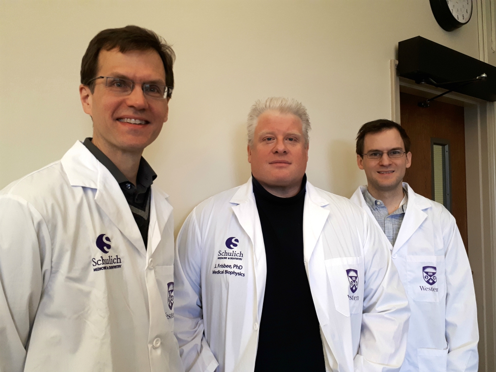
Medical Biophysics NSERC Winners
 Corey Baron, Ph.D.
Program Title: Diffusion MRI at Ultra-High Field
Diffusion magnetic resonance imaging (MRI) measures parameters describing water diffusion (i.e. random molecular motion) in tissue. Diffusion is strongly affected by cell membranes, which allows diffusion MRI to non-invasively probe microscopic tissue characteristics. Accordingly, diffusion MRI has provided a unique ability to study changes in the brain that occur during development and in disease.
Diffusion MRI at ultra high fields increases the signal strength; however, diffusion MRI suffers from severe distortions as magnetic field strength is increased. Some progress towards solving this distortion issue has been made, but these methods drastically increase scan time. The objectives of this proposal are to develop methods for distortion-free and time-efficient diffusion MRI at ultra high fields. This work will involve the design of new data sampling schemes and exploitation of data redundancies to reduce distortions and decrease scan times, and the development of “model-based reconstructions” that to remove any remaining distortions. Accordingly, this work will open the door to a whole new set of neurological investigations in these regions, both at ultra high field and normal field strengths.
Keith St. Lawrence, Ph.D.
Program Title: Developing Optical Methods to Study Dynamic Blood Flow/Metabolism Regulation in the Brain
The brain uses more energy than any other organ in the human body, accounting for a staggering 20% of the daily energy intake despite weighting less than 2% of the body’s weight. In addition, the brain has no energy stores so it relies on fine tuning of blood flow to deliver a continuous supply of fuel (i.e., oxygen and glucose). Adjustments to blood flow occur regionally throughout the brain as local energy consumption is constantly changing to match variations in brain activity. This tight coupling between blood flow, energy consumption and brain activity has led to the recognition that proper control of blood flow is critical to brain health. Disease states such as stroke or Alzheimer’s disease can reflect changes to blood flow control hindering adequate delivery of oxygen and glucose in response to local brain activity.
To better how understand this dynamic process, this research program is focused on developing a light-based device to measure the coupling of blood flow to energy consumption during changes in brain activity. The technology is extremely safe, inexpensive, and versatile (i.e. brain activity can be monitored during any number of activities, such as exercise). Our goal is to combine two light-based methods that provide complementary information of brain function: one is sensitive to oxygen use, while the other measures blood flow. It is our vision that these methods working together will provide a rich data set to begin understanding how subtle changes in blood flow control can impact the brain’s energy needs and if these changes can be linked to possible age-related issues such as cognitive decline.
Jefferson Frisbee, Ph.D.
Program Title: “Vascular Integration: How do resistance arterioles merge multiple vasomotor inputs for a coordinated response?”
Resistance arterioles play a critical role in regulating bulk blood flow to, and perfusion distribution within, tissues and organs. To do this effectively, they must integrate a myriad of vasomotor stimuli in a manner that allows them to regulate their tone effectively and produce an appropriate outcome. While there has been extensive investigation on the major individual regulators of vascular tone (e.g., myogenic activation, adenosine-induced dilation), there has been comparatively little investigation into how these stimuli are integrated by the arteriole to produce the outcome. To address this, we will use both novel and established multi-scale techniques for studying vascular function, to the question of parameter interaction for vascular tone regulation.
Microvascular networks will be studied using in situ skeletal muscle preparations, while ex vivo microvessels will be assessed using established cannulation procedures. To gain superior multi-scale validity, we will employ established in vivo hindlimb preparations to gain insight into integrated microvascular network and resistance arteriolar behavior. Integrating across scales, we incorporate novel quantitative approaches to identify critical targets for interrogation.


